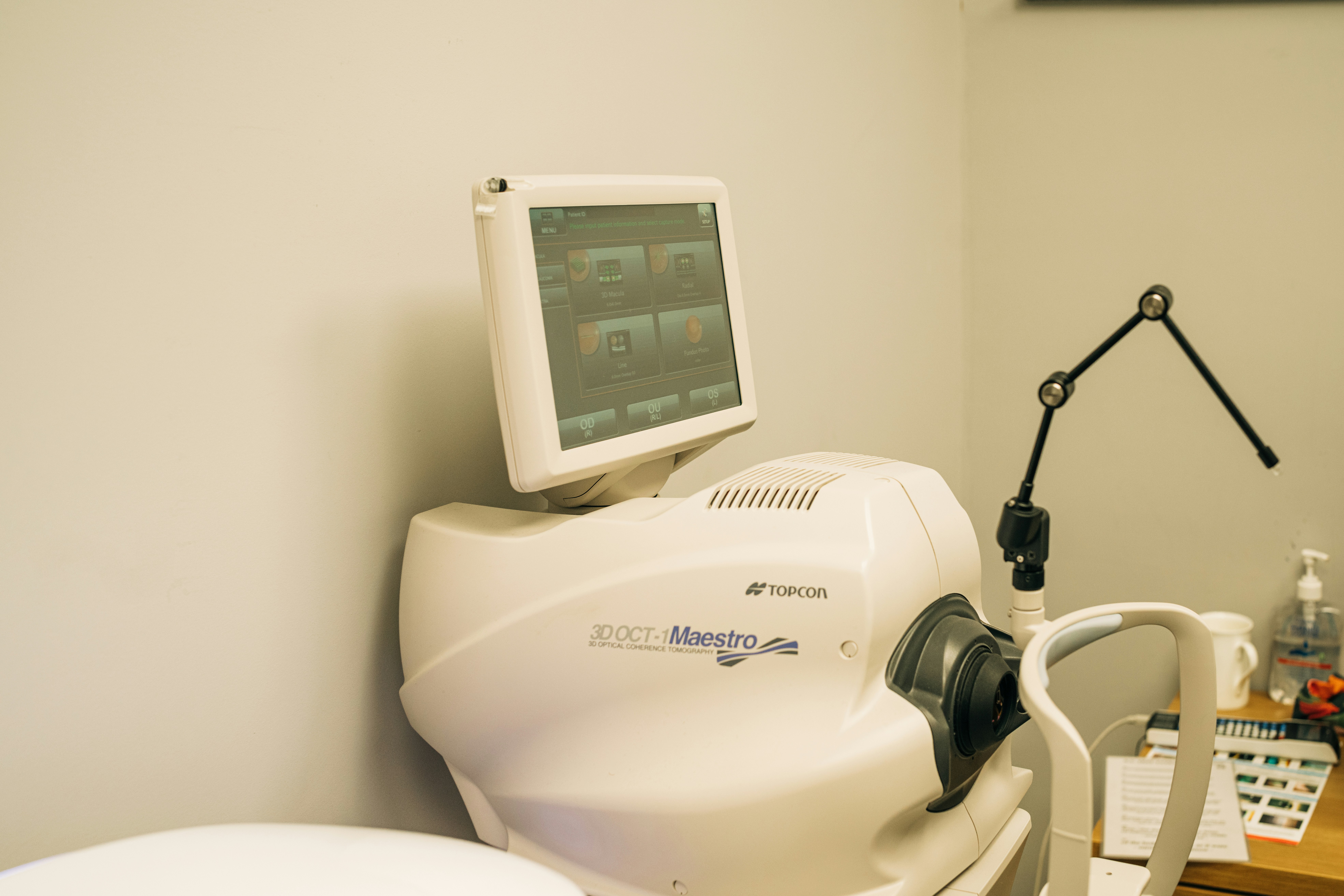
Introduction to High-End Radiology Equipment
High-end radiology equipment represents a pivotal advancement in the field of modern medicine, significantly enhancing diagnostic capabilities and treatment strategies. Radiology, the branch of medicine that employs imaging techniques to diagnose and treat diseases, relies heavily on sophisticated machinery. These advanced tools not only improve the accuracy of diagnoses but also streamline treatment planning and enhance overall patient care. The evolution of radiology equipment has transformed it from basic imaging technologies to highly specialized machines that deliver detailed insights into a patient’s health.
The significance of high-end radiology equipment cannot be overstated. Sophisticated imaging technologies such as Magnetic Resonance Imaging (MRI), Computed Tomography (CT) scans, and Positron Emission Tomography (PET) scans provide invaluable information that assists healthcare providers in identifying medical conditions at earlier stages. These early detections contribute to more effective treatment options, reduced hospital stays, and improved health outcomes. Additionally, high-end radiology equipment plays a crucial role in ongoing monitoring of patients during their treatment journeys, ensuring timely interventions when necessary.
Various types of radiology machines underscore the diversity and specialization within this field. For instance, an MRI machine utilizes powerful magnets and radio waves to produce detailed images of organs and tissues, making it essential for neurological and musculoskeletal evaluations. Conversely, CT scanners combine multiple X-ray images to generate cross-sectional views of internal structures, aiding in the diagnosis of various conditions ranging from cancer to internal injuries. Beyond these, ultrasound machines use sound waves to visualize soft tissues, serving as a non-invasive option for many diagnostic procedures.
As we explore the evolution and importance of high-end radiology equipment, it becomes evident that these machines not only support diagnostic accuracy but also embody the future of personalized medicine. The ongoing advancements in technology promise to further enhance the capabilities of radiology, ensuring that it continues to meet the ever-evolving needs of patients and healthcare providers alike.
Linear Accelerators: Precision in Radiation Therapy
Linear accelerators, commonly referred to as LINACs, represent a significant advancement in the field of radiation therapy for cancer treatment. These sophisticated machines are designed to deliver high-energy X-rays or electrons precisely to malignant tumors while minimizing exposure to surrounding healthy tissue. The technology behind LINACs utilizes accelerated electrons, which are generated through microwave technologies and subsequently directed towards a target to produce radiation. This targeted approach is crucial in treating various types of cancer effectively.
The advantages of using LINACs over traditional treatment methods, such as orthovoltage therapy, are notable. LINACs enable precision targeting of tumors, which allows for higher doses of radiation to be administered directly to the cancerous cells, thereby increasing treatment efficacy. Additionally, their ability to deliver radiation in three dimensions, often accomplished through techniques such as Intensity-Modulated Radiation Therapy (IMRT) and Volumetric Modulated Arc Therapy (VMAT), significantly enhances the capability to create individualized treatment plans tailored to each patient’s anatomy.
LINACs are not only integral to delivering effective radiation treatments but also play a vital role in comprehensive cancer care. The integration of these advanced machines into treatment protocols ensures that patients receive holistic care along with ongoing advancements in imaging technology, such as PET and CT scans, which facilitate precise localization of tumors prior to treatment. Furthermore, continuous innovations in LINAC technology, including motion management systems and adaptive therapy modes, have improved patient safety and comfort during procedures.
To ensure patient safety, numerous measures are implemented during the use of linear accelerators. This includes routine calibration and maintenance protocols, as well as the development of protocols for emergency situations. Such practices are critical in promoting a secure environment for both patients and staff, reinforcing the significance of LINACs in modern oncology treatment.
Magnetic Resonance Imaging (MRI): Unveiling the Human Body
Magnetic Resonance Imaging (MRI) has revolutionized the field of diagnostic imaging by offering exceptionally detailed images of soft tissues and organs. Unlike traditional imaging technologies such as X-rays or CT scans, MRI utilizes strong magnetic fields and radio waves to generate detailed cross-sectional views of the human body, which are invaluable for medical diagnoses. This non-invasive technique excels in visualizing the anatomy and physiology of soft tissues, making it indispensable in various medical specialties.
The underlying principle of MRI technology is based on the magnetic properties of hydrogen nuclei present in the body. When a patient is placed in a strong magnetic field, the hydrogen atoms align with the magnetic field. Radio frequency pulses are then applied, causing these atoms to emit signals that are detected and converted into images by sophisticated computing algorithms. The resulting images provide remarkable clarity and detail, which can be manipulated to view structures from multiple angles.
There are several types of MRI scans, including functional MRI (fMRI), which measures brain activity by detecting changes in blood flow, and contrast-enhanced MRI, which uses contrast agents to improve the visibility of certain tissues. Each type has unique applications; for instance, fMRI is commonly used in neurological studies, while contrast-enhanced MRI plays a crucial role in oncology for identifying tumors.
MRI offers several significant benefits over other imaging modalities. Its ability to produce high-resolution images without exposing patients to ionizing radiation is a notable advantage, making it a safer option for frequent use. Additionally, MRI can capture images in real-time, facilitating dynamic studies of organ function, particularly in the cardiovascular system and the brain. Overall, MRI continues to be a vital tool in modern medicine, enabling healthcare providers to diagnose and manage a wide range of medical conditions effectively.
Computed Tomography (CT) Scans: A Cross-Sectional View
Computed Tomography (CT) scans have revolutionized the field of diagnostic imaging, providing detailed cross-sectional views of the human body. Utilizing advanced computer algorithms and x-ray technology, CT scans create high-resolution images that allow healthcare professionals to visualize and evaluate internal structures with unprecedented clarity. Each scan involves rotating x-ray sources around the patient, capturing multiple images from various angles. These images are then digitally processed to generate comprehensive 2D and 3D representations of the examined area.
Various applications of CT scans are noteworthy in clinical practice. They play a pivotal role in evaluating traumatic injuries, detecting tumors, mapping out diseases, and guiding interventions like biopsies. Specific types of CT scans include angiography, which focuses on blood vessels, and high-resolution scans aimed at identifying lung anomalies. The versatility of CT technology further expands into specialized domains such as cardiac CT, which aids in assessing coronary artery disease, or dental CT, essential for imaging complex dental structures.
Recent advancements in CT scanning technology have enhanced accuracy and reduced scan time, with innovations such as 3D reconstruction offering detailed views of anatomical structures. This capability assists in surgical planning and patient consultations, allowing surgeons to visualize the anatomy before performing procedures. However, the use of CT scans is not without considerations. Radiation exposure remains a concern, prompting ongoing research into minimizing dosages while maximizing diagnostic efficacy. The implementation of dose-reduction software and the development of new technologies, such as iterative reconstruction techniques, have significantly improved safety profiles without compromising image quality.
In conclusion, CT scans are an invaluable part of modern medicine, providing a cross-sectional view that underscores their significance in effective diagnosis and treatment protocols, while also posing challenges that necessitate careful consideration of patient safety and innovation in technology.
The Role of X-Ray Machines in Diagnostics
X-ray machines have long been a cornerstone in the field of medical diagnostics, providing crucial insights through the visualization of the internal structures of the human body. Traditional X-ray technology operates on the principle of radiography, where controlled doses of ionizing radiation are directed toward a patient’s body. As these rays pass through, they are absorbed varying levels by different tissues, with denser structures like bones appearing white on the X-ray film, while softer tissues appear darker. This distinct contrast allows clinicians to diagnose a variety of conditions, including fractures, infections, and various diseases.
The evolution of X-ray machines from analog to digital systems has significantly benefited the field. Traditional X-ray machines utilized film that required careful developing processes, often resulting in longer wait times for diagnostic results. However, with advancements in digital X-ray technology, images are now processed electronically, drastically improving efficiency and image quality. Digital X-ray systems use sensors to capture images, providing immediate access to high-resolution pictures that can be easily manipulated and enhanced for better interpretation.
The applications of X-ray machines in modern medicine extend beyond mere fracture detection. They are instrumental in diagnosing conditions such as pneumonia, tumors, and even dental issues through specialized radiographic techniques. The increased precision and reduced radiation exposure associated with digital X-rays have been pivotal in enhancing patient safety. Furthermore, improved storage and sharing capabilities of digital images facilitate collaboration among healthcare professionals, streamlining the diagnostic process.
In conclusion, the advancements in X-ray machines have profoundly transformed diagnostic imaging, making them an integral part of modern medicine. Their ability to provide swift, accurate diagnostics continues to underscore their importance in various healthcare settings.
Gamma Cameras: Imaging in Nuclear Medicine
Gamma cameras are pivotal instruments in the realm of nuclear medicine, renowned for their capacity to visualize metabolic processes within the human body. Utilizing a technique that involves the detection of gamma radiation emitted from radioactive tracers administered to patients, these cameras produce detailed images that elucidate functional activity in organs and tissues. This non-invasive imaging modality significantly enhances diagnostic accuracy and plays a crucial role in managing various medical conditions.
The operation of a gamma camera involves the meticulous integration of several components, including a collimator, scintillation crystal, and photomultiplier tubes. When a radioactive tracer is injected into the patient, it emits gamma rays which are captured by the gamma camera. The collimator ensures that only those rays traveling in specific directions reach the scintillation crystal. Subsequently, the crystal converts the gamma radiation into visible light, which is then transformed into electrical signals by photomultiplier tubes. These signals are processed to construct clear images representing metabolic activity, allowing healthcare professionals to interpret physiological changes and diagnose ailments effectively.
Gamma cameras find extensive application across various medical disciplines, particularly in cardiology, oncology, and neurology. In cardiology, they are instrumental in evaluating cardiac function through techniques such as myocardial perfusion imaging. In oncology, they assist in detecting tumors and monitoring treatment responses by visualizing the uptake of radiotracers in malignant tissues. Additionally, in neurology, gamma cameras facilitate the assessment of brain disorders such as Alzheimer’s disease by monitoring metabolic activity. The evolving integration of gamma cameras with other advanced imaging modalities, such as CT or MRI, enhances diagnostic capabilities, providing comprehensive insights into patient health.
Ultrasound: A Non-Invasive Diagnostic Tool
Ultrasound technology has become a cornerstone in the field of modern medicine, offering a non-invasive way to visualize internal structures of the body. By utilizing high-frequency sound waves, ultrasound creates real-time images that can be crucial for diagnosing a variety of medical conditions. This technology has found substantial applications in diverse medical fields, including obstetrics, cardiology, and abdominal imaging.
In obstetrics, ultrasound is routinely employed to monitor fetal development, assess gestational age, and identify potential complications during pregnancy. The ability to visualize a developing fetus non-invasively has revolutionized prenatal care, allowing healthcare providers to ensure both maternal and neonatal well-being. Additionally, ultrasound technology has developed advanced features such as 3D imaging, enabling more detailed examination of fetal anatomy.
In cardiology, ultrasound, particularly Doppler ultrasound, facilitates the evaluation of cardiac structures and blood flow. This non-invasive technique allows cardiologists to diagnose various heart conditions, including valve disorders and congenital defects. The credibility of Doppler ultrasound has made it an integral part of cardiac assessments, enabling precise measurements of blood flow velocities and identifying abnormalities in circulation.
Furthermore, abdominal imaging has greatly benefited from advances in ultrasound technology. It serves as a valuable tool in assessing organs such as the liver, kidneys, and gallbladder. Non-invasive abdominal ultrasound can help detect conditions like gallstones, liver diseases, and tumors, replacing more invasive procedures that carry higher risks and complications.
The evolution of ultrasound technology continues to enhance its capabilities and applications in medicine. Innovations, including high-resolution transducers and advanced image processing software, have improved diagnostic accuracy, making it an invaluable tool in patient care. As the technology progresses, the breadth of ultrasound applications expands, ensuring its pivotal role in modern medical diagnostics.
Angiography: Visualizing Blood Vessels
Angiography is a crucial imaging technique in modern medicine, particularly for visualizing blood vessels and diagnosing a range of vascular disorders. This method employs X-ray technology, often coupled with a contrast agent, to create detailed images of the blood vessels, allowing for the accurate assessment of conditions such as blockages, aneurysms, and other vascular abnormalities.
There are several types of angiography, with coronary and cerebral angiography being among the most common. Coronary angiography focuses on the coronary arteries that supply blood to the heart. This technique is vital in diagnosing coronary artery disease, where narrowed or blocked arteries can lead to serious complications such as heart attacks. In contrast, cerebral angiography targets the blood vessels in the brain, aiding in the evaluation of conditions like strokes, arteriovenous malformations, and brain tumors.
The process of catheterization, a key aspect of angiography, involves inserting a thin, flexible tube (catheter) into a blood vessel, typically through the wrist or groin. The catheter is threaded through the vascular system to the area of interest, where contrast material is injected. This substance absorbs X-rays more than the surrounding tissues, allowing for high-contrast images that highlight vascular structures. The visualization that angiography provides not only assists in diagnosis but also enables interventional procedures, such as balloon angioplasty and stent placement, where appropriate.
Recent advancements in angiography technology have greatly improved the efficacy and safety of these procedures. Innovations such as digital subtraction angiography (DSA) enhance image clarity by subtracting non-contrast images, allowing for a clearer view of the vessels. Furthermore, the integration of three-dimensional imaging and real-time monitoring has revolutionized how vascular conditions are evaluated and treated. Collectively, these advances underscore the importance of angiography in patient care, facilitating timely and accurate interventions that can significantly improve outcomes.
PACS and Laboratory Analyzers: Enhancing Workflow and Diagnosis
The integration of Picture Archiving and Communication Systems (PACS) into modern healthcare has revolutionized the way medical images are stored, managed, and shared. By facilitating the digital storage of imaging data, PACS eliminates the need for cumbersome physical imaging materials, thereby ensuring greater efficiency in workflow and significantly improving the management of radiological resources. High-end radiology equipment, such as MRI and CT scanners, seamlessly aligns with PACS, resulting in streamlined processes from image acquisition to interpretation. This integration not only makes access to critical diagnostic information instantaneous but also enhances collaboration among healthcare professionals, ultimately leading to improved patient care outcomes.
PACS enhances workflow by allowing medical personnel to quickly retrieve images and reports from a centralized database. This immediacy aids radiologists and other specialists in making faster, more informed diagnostic decisions. The capability to view and analyze images from remote locations further reduces delays in treatment, particularly in emergency situations where time is of the essence. Furthermore, with the implementation of advanced technologies, such as cloud-based PACS solutions, healthcare facilities can achieve greater scalability and efficiency, accommodating fluctuating image storage needs without compromising on speed or accessibility.
In addition to PACS, laboratory analyzers play a crucial role in diagnostics across various medical specialties. These high-end instruments, including hematology analyzers, biochemistry analyzers, and immunoassay analyzers, are vital for conducting lab tests that are fundamental to effective patient diagnosis and management. By automating the analytical processes, these devices not only enhance the accuracy of results but also drastically reduce the turnaround time for test outcomes. The integration of laboratory analyzers with information systems further streamlines laboratory operations, leading to optimal resource allocation and improved overall healthcare delivery. The combined advancements in PACS and laboratory analyzers underscore their critical importance in modern medicine, highlighting their roles in enhancing diagnostic efficiency and patient care.
Hybrid Operating Room Systems: A Revolution in Surgical Procedures
Hybrid operating room systems represent a significant advancement in the capabilities of modern surgical practices. These sophisticated environments amalgamate traditional surgical equipment with advanced imaging technologies, such as computed tomography (CT) and magnetic resonance imaging (MRI), to foster real-time visualization of anatomical structures during procedures. This unique integration enhances the precision of surgeries, ultimately leading to improved patient outcomes and greater safety.
The core components of a hybrid operating room typically include high-definition imaging systems, surgical tables designed for optimal patient positioning, and advanced lighting systems that facilitate visibility. Surgeons can use various imaging modalities without needing to move the patient, allowing for continuous assessment of their conditions during surgical interventions. This immediate access to imaging data not only aids in the initial diagnosis but also assists in making real-time decisions based on the evolving surgical landscape.
One of the main benefits of employing hybrid operating room systems is the enhancement of minimally invasive surgical techniques. Procedures such as vascular surgeries and cardiac interventions greatly benefit from real-time imaging, as surgeons can navigate complex anatomy with greater accuracy. Minimally invasive techniques often result in reduced recovery times, less postoperative pain, and shorter hospital stays, underscoring the importance of these advanced systems in modern surgical practice.
As technology evolves, the future of hybrid operating room systems appears promising. The ongoing development of more compact, efficient imaging devices and augmented reality solutions is likely to further enhance the scope of procedures conducted in these settings. The integration of artificial intelligence and machine learning may also allow for predictive analytics to support surgical decision-making and outcomes, marking a leap forward in surgical techniques. Overall, hybrid operating room systems are poised to play a crucial role in the continual advancement of surgical practices, ultimately benefiting patients worldwide.


 Healthcare Technology